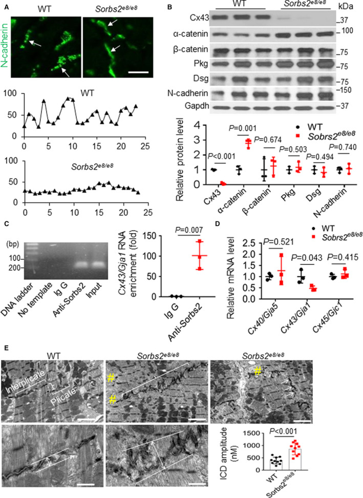
Figure 5. Dual function of Sorbs2 (sorbin and SH3 domain‐containing 2) in maintaining intercalated disc (ICD) integrity and regulating the expression of α‐catenin and gap junction protein connexin 43 (Cx43).

A, Shown are ICD images and represented pseudoline analysis of mouse hearts after immunostaining using an anti–N‐cadherin antibody. The expression of N‐cadherin in ICD manifests as discontinuous dots in wild‐type (WT) mice, which switched to continuous lines in Sorbs2e8/e8 mice (arrows). Bar=20 μm. The intensity distribution of the N‐cadherin signal along the ICD appears as wavy lines in WT mice, which largely changed to flat lines in the Sorbs2e8/e8 mice. B, Western blotting and quantification of ICD proteins in WT and Sorbs2e8/e8 knockout (KO) mice hearts at 4 months old. N=3. Unpaired 2‐tailed Student t test. C, A representative DNA gel and quantitative reverse transcription–polymerase chain reaction (RT‐PCR) analysis of the Cx43/Gja1 mRNA level in the anti‐Sorbs2 antibody and IgG control immunoprecipitated tissue lysates from right ventricle (RV) of WT mice. N=3. Unpaired 2‐tailed Student t test. D, Quantitative RT‐PCR analysis of the mRNA levels of the indicated genes in the RV tissue of Sorbs2e8/e8 mice compared with WT controls. N=3. Unpaired 2‐tailed Student t test. E, Transmission electron microscopy images and schematic illustration on measuring and quantifying the ICD amplitude in WT and Sorbs2e8/e8 KO mice hearts at 4 months old. #Disrupted myofibril. Bars for top panels=2 μm; bars for bottom panels=500 nm. N=10. Unpaired 2‐tailed Student t test. Dsg indicates desmoglein 1/2; and Pkg, plakoglobin.
