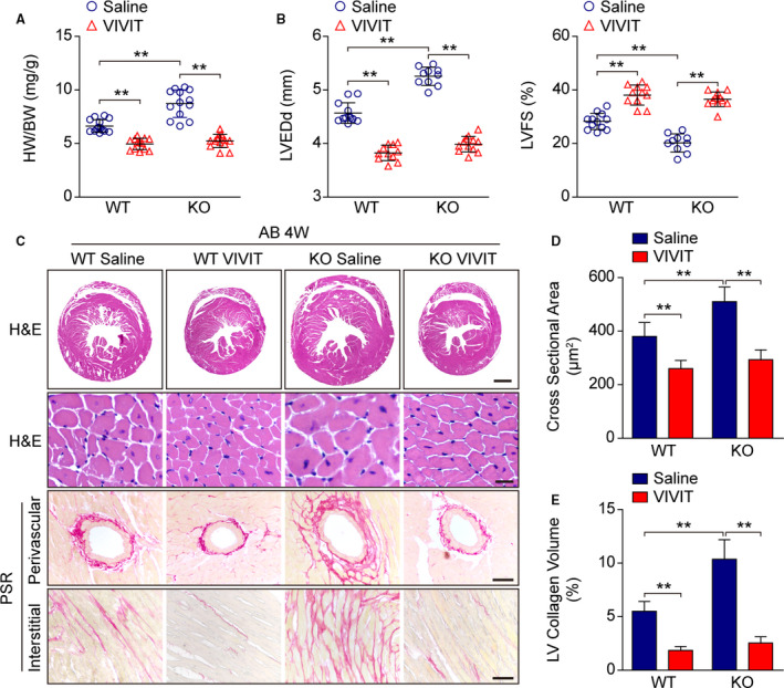
Figure 7. Nuclear factor of activated T cells (NFAT) inactivation rescues the aggravated pathology by Nulp1 deletion in vivo.

A, Comparison of heart weight (HW)/body weight (BW) ratios in wild‐type (WT) and Nulp1 knockout (KO) mice subjected to 4 weeks of aortic banding (AB) surgery with or without NFAT inhibitor (VIVIT) treatment. n=13 to 14 mice per group. B, Echocardiographic parameters left ventricular end‐diastolic diameter (LVEDd) and left ventricular fractional shortening (LVFS) in the indicated groups. n=10 to 12 mice per group. C, Histological analyses by hematoxylin and eosin (H&E) staining of whole‐heart short‐axis cross sections (the first row; scale bar, 1000 μm) and individual myofibril cross sections (the second row; scale bar, 50 μm) and representative images of cardiac fibrosis, as visualized by picrosirius red (PSR) staining of the perivascular area (the third row; scale bar, 50 μm) and interstitial area (the fourth row; scale bar, 50 μm) in the indicated groups. n=6 to 8 mice per group. D, Results pertaining to the cross‐sectional areas of the cardiomyocytes in the indicated groups (n≥100 cells per group). E, Quantification of LV collagen volumes in the indicated groups (n≥40 fields per group). For all statistical plots, the data are presented as the mean±SD; in (A, B, D, and E) **P<0.01; the statistical analysis was performed using a 1‐way ANOVA.
