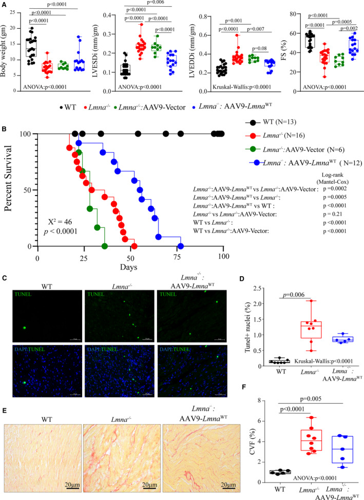
Figure 6. Phenotypic consequences of AAV9 mediated LmnaWT expression in the Lmna−/− mice.

A, Selected echocardiographic indices of cardiac structure and function showing improvement of cardiac function upon vector or AAV9‐Lmna WT injection of Lmna−/− mice at 4 weeks after birth. LVEDDi: Left Ventricular End‐Diastolic Diameter indexed to the bodyweight; LVESDi, left ventricular end‐systolic diameter indexed to the bodyweight; FS, fractional shortening (age, sex, and number of mice used in each group are listed in Tables S3 and S7). B, Kaplan–Meier survival plots of WT, Lmna−/−, and Lmna−/− mice injected with vector or AAV9 expressing LmnaWT. Chi‐square and P value for the overall Kaplan–Meier survival analysis are indicated in bold. Log‐rank (Mantel‐Cox) pairwise analysis P values for each subgroup analysis are shown. C, Representative terminal deoxynucleotidyl transferase deoxyuridine triphosphate nick end labeling (TUNEL) stained thin myocardial cross‐section from 4‐week‐old WT, Lmna−/−, and Lmna−/−: AAV9‐Lmna WT are shown. The lower panel shows overlay images of TUNEL staining in green and nuclei (blue). D, Respective quantitative data of the TUNEL‐positive stained nuclei in WT (n=7), Lmna−/− (n=8), and Lmna−/− mice injected with AAV9‐Lmna WT (n=5). E, Representative Picrosirius red‐stained thin myocardial sections from 4‐week‐old WT, Lmna−/−, and Lmna−/−: AAV9‐Lmna WT. F, Respective quantitative data on collagen volume fraction in WT (n=5), Lmna−/− (n=8), and Lmna−/− mice injected with AAV9‐Lmna WT (n=5). One‐way ANOVA followed by Tukey’s multiple pairwise comparison P values is shown. Only P values that were significant (P<0.05) are shown. AAV9 indicates adeno‐associated virus serotype 9; KMD5, lysine‐specific demethylase 5; and WT, wild‐type.
