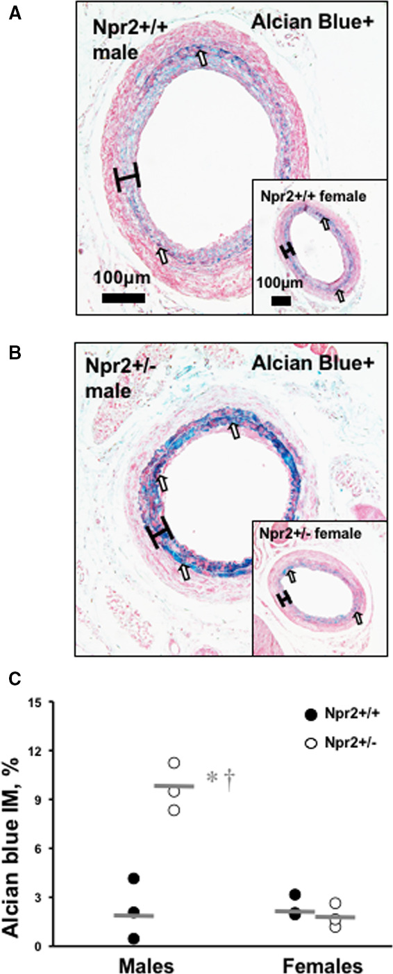
Figure 5. Differences in carotid fibrosis in Npr2 mice.

A, Representative image of PicroSirius Red–stained ligated LCA in male Npr2 wild‐type (Npr2 +/+) mouse. B, Representative image of Alcian Blue–stained ligated LCA in male Npr2 heterozygous (Npr2 +/−) mouse. Insets show corresponding females. Scale bar=100 μm. Black brackets indicate intima/media area. C, Quantification of fibrosis (blue color) in intima/media area of the LCA (%). Black circles indicate individual Npr2 +/+ mice. Open circles indicate Npr2 +/− mice. Gray lines indicate mean values. *P<0.001 vs Npr2 +/+ males; † P<0.001 vs Npr2 +/− females. n=3 animals per group. LCA indicates left carotid artery.
