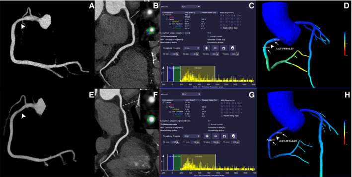
Figure 4. Representative case of a 77‐year‐old woman showing similar △computed tomography‐fractional flow reserve after statin treatment.

A through D, The baseline coronary computed tomography angiography revealed calcified lesion with mild stenosis of proximal left anterior descending (white arrowhead). The total plaque volume was 53.56 mm3 and △computed tomography‐fractional flow reserve of this lesion was 0.07. E through H, The follow‐up coronary computed tomography angiography (14 months later) after statin treatment showed mild stenosis of proximal left anterior descending (white arrowhead). The total plaque volume was 53.77 mm3 and follow‐up △computed tomography‐fractional flow reserve was 0.06, which were both similar to baseline measurements. CCTA indicates coronary computed tomography angiography; CT, computed tomography; FFR, fractional flow reserve; LAD, left anterior descending; and TPV, total plaque volume.
