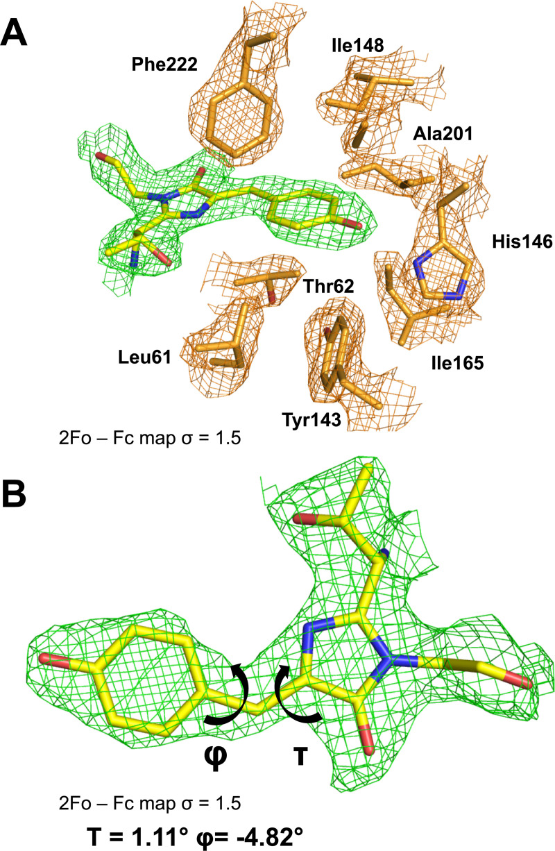
Fig 5. The AausFP1 chromophore environment.
(A) 2Fobs − Fcalc electron-density map contoured at a 1.5 σ level superimposed over the model of the chromophore and the neighboring residues in the structure of AausFP1. (B) Dihedral angle definition around the chromophore methylene bridge. The data underlying this figure may be found in PDB 6S67.

