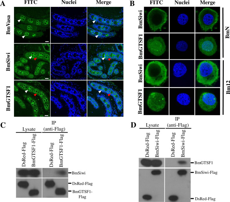
Fig 6. BmGTSF1 interacts with BmSIWI.
(A) Localization of BmVasa, BmSIWI and BmGTSF1 in the ovaries of B. mori. A FITC-conjugated secondary antibody was used for fluorescence detection and Hoechst staining (blue) showed the locations of the nuclei. Scale bar, 50μm. The white and red arrow indicates germ line cells and somatic cells respectively. (B) Intracellular localization of BmSIWI and BmGTSF1 in BmN cells and Bm12 cells. (C) Immunoprecipitation followed by western blot showed that endogenous BmSIWI immunoprecipitated with BmGTSF1-Flag rather than DsRed-Flag. (D) Immunoprecipitation followed by western blot showed that endogenous BmGTSF1 immunoprecipitated with BmSIWI-Flag rather than DsRed-Flag.

