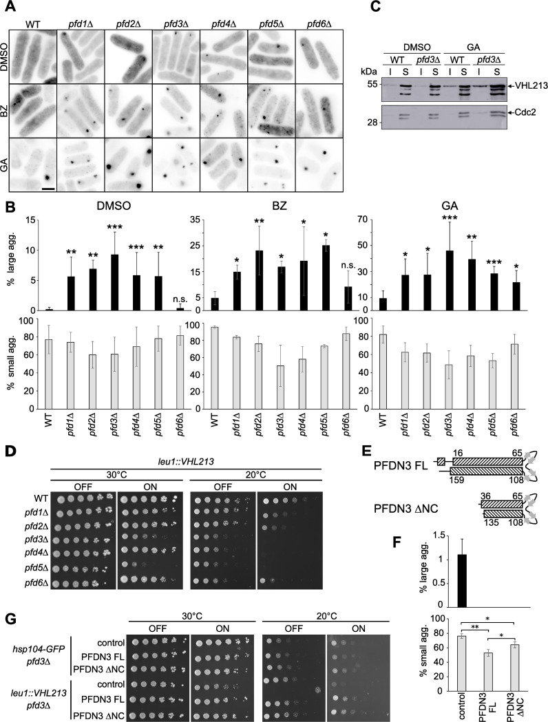
Fig 4. Aggregation and toxicity of pVHL213 are increased in prefoldin mutants.
Cells of leu1::VHL213 (WT) and leu1::VHL213 prefoldin mutant as indicated were expressing GFP-pVHL213. A) Reversed fluorescent images of the different strains expressing GFP-pVHL213 treated with DMSO, BZ or GA. Bar: 5 μm. B) Histograms representing the percentage of large (upper panels) and small (lower panels) aggregate-containing cells in DMSO, BZ and GA treated cells (mean±s.d. from at least three independent experiments, *, p<0.05; **, p<0.01; ***, p-value<0.001; WT vs pfd mutant for each condition, Mann-Whitney test), C) Western blot analysis of GFP-pVHL213 expression in WT and prefoldin leu1::VHL213 pfd3Δ mutant in the presence of GA or DMSO as a control: Soluble (S) and Insoluble (I) fractions are shown. Cdc2 was used as a loading control. D) Serial dilutions (1:5) of the indicated strains were spotted on EMM plates in the OFF or ON conditions and incubated for 5 days at 30°C and 7 days at 20°C as indicated. E) Scheme representing the secondary structures of full-length (FL) or mutated (ΔNC) human PFDN3 protein: α-helices and β-strands are represented by dashed boxes and grey arrows, respectively. Numbers refer to the amino acids in the full-length PFDN3 protein. F) Histograms representing the percentage of large (upper panel) and small (lower panel) aggregate-containing cells in leu1::VHL213 pfd3Δ cells after expression of the full-length (FL) or mutated (ΔNC) human PFDN3 protein or in control cells transformed with empty vector (mean±s.d. from three independent experiments), G) Serial dilutions (1:5) of the indicated strains were spotted on EMM plates in the OFF or ON conditions and incubated for 5 days at 20°C and 30°C as indicated. Hsp104-GFP pfd3Δ transformants were used as a wild-type control.

