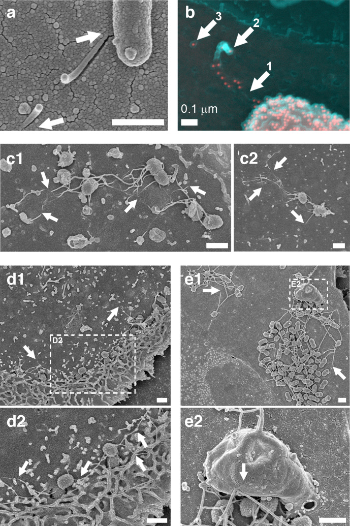
Fig. 4.
SEM of E. coli O157:H7 and S. Typhimurium flagella-like structures disappearing and reappearing at host cell surfaces. (a, b) E. coli O157 : H7 (ZAP734) interactions with bovine primary epithelial cell cultures 3 h post-infection. (a) Secondary electron (SE) image of a snapped flagella-like filament, which appears to penetrate the cell surface (arrows). (b) A false-coloured SE image (cyan) superimposed with a false-coloured back-scattered image (red). H7 flagella and O157 LPS are both immuno-gold labelled. Filament staining occurs adjacent to the bacterium and is then absent (arrow 1). The filament is then broken and curls back on itself (arrow 2), with the remnant embedded filament (arrow 3). (c1, c2) SE images of flagella-like filaments disappearing into and coming out of the surface of IPEC-J2 epithelial cells (arrows) within the first 30 min of infection with S. Typhimurium (Maskan). (d) SE image of S. Typhimurium (Maskan) micro-colonies on IPEC-J2 cells 40 min post-infection. (d1) Actin ruffling proximal to invading Salmonella . (d2) Enlarged from the inset indicated in (d1). Wavy flagella-like filaments are interacting with ruffled and unruffled cell surfaces (arrows). (e) SE image of S. Typhimurium SL1344 (WT) micro-colonies on bovine primary epithelial cells. (e1) Long filaments disappearing into the cell surface (arrows). (e2) Higher resolution image of the area indicated in (b1), which shows long filaments interacting with a large macropinocytic protrusion (arrow). Samples were fixed in 3 % (w/v) glutaraldehyde and were not permeabilized prior to sample processing for SEM. Imaging was undertaken on a Hitachi 4700 field emission scanning electron microscope. Scale bars, 1 µm unless indicated otherwise.

