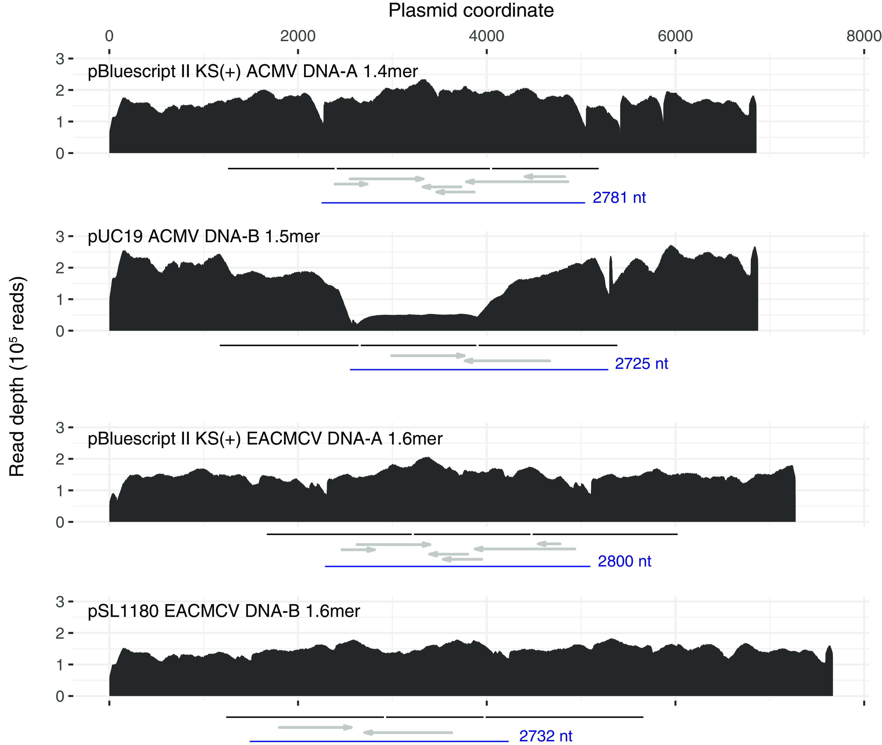
FIG 1.

Plots of Illumina read depth across the length of the four infectious clone plasmids. One of three libraries is shown for each plasmid (Sequence Read Archive accession numbers SRR12354432, SRR12354427, SRR12354424, and SRR12354421). The region in each plasmid corresponding to each virus segment partial tandem dimer unit is indicated with a black line under each graph. Vertical white lines demarcate the boundaries of the unique and duplicated regions of each concatemer. Each virus segment monomer unit (between two replication origin nick sites) is shown in blue. Canonical virus genes are indicated with gray arrows, left to right for virus sense (AV1, AV2, and BV1) and right to left for complementary sense (AC1 to AC4 and BC1). The uneven read depth for the ACMV DNA-B plasmid is due to instability (truncation), which is evident in single-cut restriction digests (not shown). Such partial deletion of tandem duplicated regions in E. coli is not uncommon (24).
