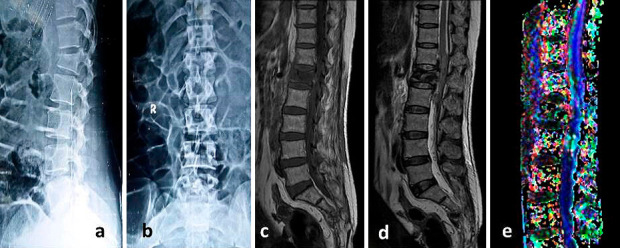
Figure 1.

Initial plain radiographs anteroposterior (a) & lateral (b) showing fracture of lumbar first vertebra in a 42 year old female with AIS (ASIA impairment scale) C neurological deficit. Conventional MRI T1-weighted (c) and T2-Weighted (d) sagittal sections shows fracture of vertebral body with spinal cord edema & hemorrhage. Fiber tractography (e) shows disruption of fibers. Values of FA, ADC and AI were 0.456, 1.42, and 0.316 respectively.
