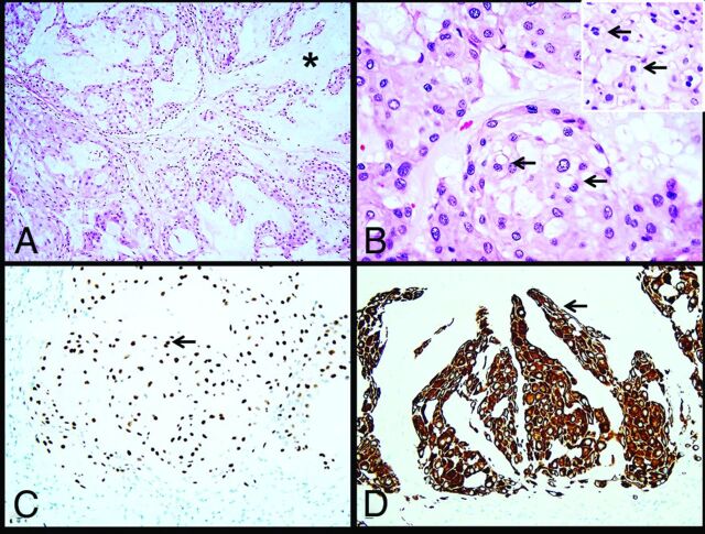
FIG 3.
Histology images. The tumor consists of cells arranged in nests and cords (A) within an abundant mucoid matrix (A, asterisk). Most of the tumor cells are large with central nuclei and mildly vacuolated eosinophilic cytoplasm (B, arrows). Scattered physaliphorous cells are also present (B inset, arrows). Immunohistochemistry demonstrates nuclear brachyury (C, arrow) and cytoplasmic keratin CAM 5.2 (D, arrow) expression in tumor cells.

