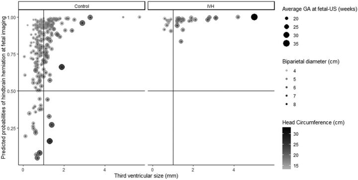
FIG 4.
Effect of third ventricular diameter, biparietal diameter, and head circumference per study group according to logistic regression results. Scatterplots depicting the effect of the model described in Table 1 for controls (left) and cases of intraventricular hemorrhage (right). Y-axis shows the predicted probability for finding prenatal hindbrain herniation (horizontal line indicates the probability of 50%). The x-axis shows the third ventricle diameter (mm). The size of the geometric point corresponds to gestational age, color intensity illustrates head circumference, and transparency shows biparietal diameter for each subject. Most subjects with intraventricular hemorrhage had a third ventricle at or above 1 mm (vertical line), hence its strong predictive effect (Table 1).

