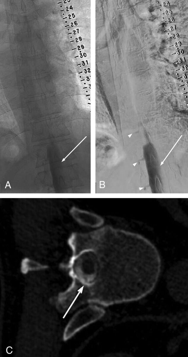
FIG 1.

Unsubtracted (A) and subtracted (B) left-side-down LDDSM images on day 1 demonstrate subdural contrast injection at the lower thoracic/upper lumbar levels. Notice nondependent masslike subdural contrast overlying the middle of the osseous spinal canal (solid arrows in A and B). Given the predominantly subdural injection, only minimal intrathecal contrast is seen filling nerve root sheaths (arrowheads in B). The study was terminated without obtaining a CTM. The next-day CTM following the repeat left LDDSM injection shows no residual subdural contrast at the level of T11 and a homogeneous appearance of the intrathecal contrast clearly outlining a nerve root traversing the intrathecal space (arrow in C). The time interval between images A/B and C is approximately 20.5 hours.
