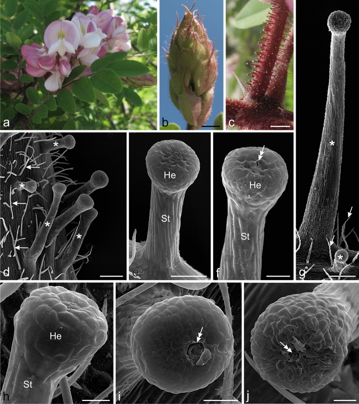
Fig. 1.
Morphology of the inflorescence and micromorphology of the glandular trichomes of Hartweg's locust. d–j SEM. a Inflorescence with flowers in different stages of development. b Visible inflorescence in the bud stage with bracts covered by glandular trichomes. c Peduncles with glandular trichomes. d Non-glandular (arrows) and massive glandular (asterisks) trichomes with varied length visible on the pedicle surface. e–j Mature glandular trichomes in different stages of activity. e, f, i, j Trichomes in the secretory or post-secretory phases with sunken cells (e, f, j) and with a pore in the cuticle (double arrows) on the head apex (f, i, j). g Non-glandular (arrows) and glandular (asterisks) trichomes with substantially varied length visible on the bract surface. h Trichome in the pre-secretory phase with convex cell walls of the head; He trichome head, St trichome stalk. Scale bars = 10 mm (b), 1 mm (c), 200 µm (d, g), 100 µm (e), 50 µm (f), 30 µm (h–j)

