Fig. 6.
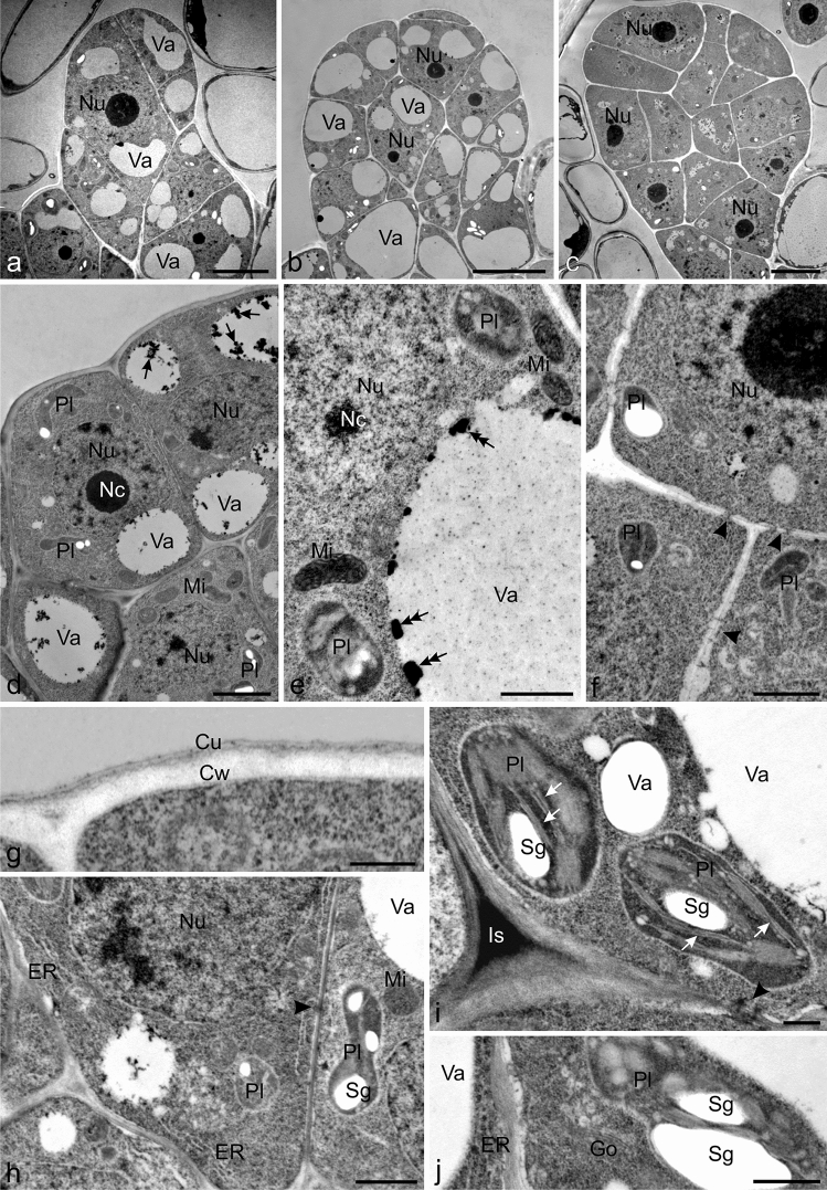
Ultrastructural traits of immature glandular trichomes of Hartweg's locust (TEM). a–c Trichomes in the initial stage of development. d Cells with a prominent nucleus with nucleoli, plastids, mitochondria, and small vacuoles with flocculent precipitates of tannins (black arrows). e Fragment of a cell with a vacuole with an electron-dense deposit of tannin near the tonoplast (double arrows) and cytoplasm with mitochondria and plastids. f Visible plastids in the cytoplasm and plasmodesmata (arrowheads) in the cell walls. g Apical fragment of a trichome with a thin cuticle on the surface. h Visible plastids with starch grains, mitochondria, ER profiles, and plasmodesmata (arrowhead) in the cell walls. i Visible plastids with starch grains and thylakoids (white arrows). j Visible plastids with starch grains, ER profiles, and Golgi apparatus with vesicles; Va vacuoles, Nu nucleus, Nc nucleoli, Pl plastids, Sg starch grains, Mi mitochondria, Pl plasmodesmata, Cw cell walls, Cu cuticle, ER endoplasmic reticulum, Is intercellular space, Go Golgi apparatus. Scale bars = 10 µm (b, c), 5 µm (a), 2 µm (d), 1 µm (e, f, h), 500 nm (g), 200 nm (i, j)

