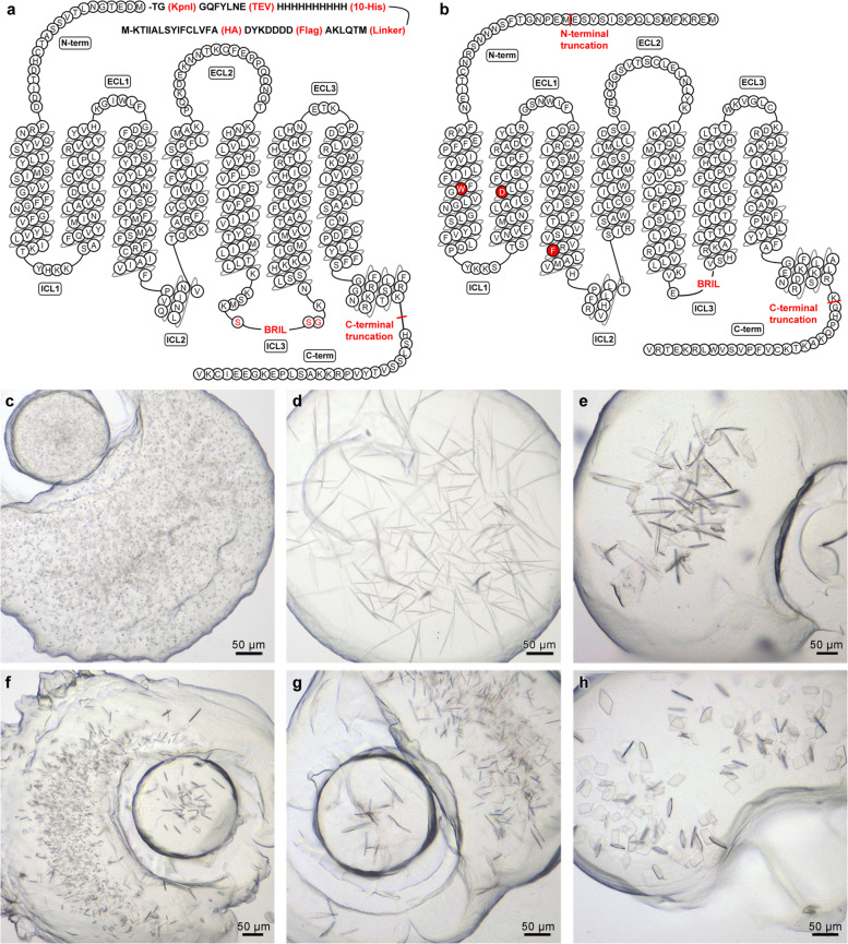
Fig. 1.
Construct design and crystallization of CysLT1R and CysLT2R. (a,b) Amino acid sequence snake-plot of the CysLT1R (a) and CysLT2R (b) crystallization constructs. Protein modifications are shown in red, red background shows stabilising point mutations, red font amino acids – linkers for BRIL insertion. Initial figure drawn using GPCRdb49. The N-terminal peptide fragment (a) was added to both constructs. (c–h) microphotographs of typical crystals grown in lipidic cubic phase (LCP) for CysLT1R-zafirlukast (c), CysLT1R-pranlukast (d), and CysLT2R with antagonists: 11a (C2221 space group) (e), 11a (F222 space group) (f), 11b (g), and 11c (h) complexes51.

