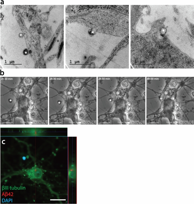
Figure 2.
Endocytosis of EVs results in intracellular Aβ deposits in cortical neurons. TEM analysis indicated that EVs attached to and were endocytosed by cortical neurons (asterisks, a). The process of EV uptake by the neurons could also be monitored with time-lapse microscopy (asterisk, b). Immunocytochemistry followed by confocal microscopy, confirmed intracellular Aβ deposits in Aβ42 protofibril-EV exposed neurons (c). Scale bars: (b) = 10 µm and (c) = 20 µm.

