Figure 4.
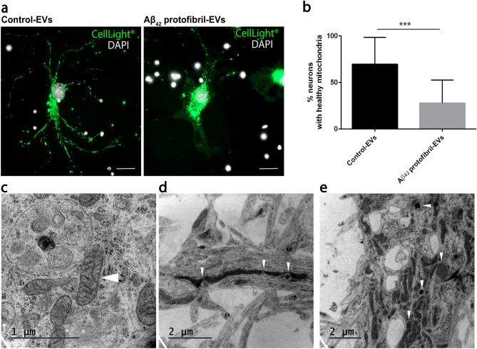
Aβ42 protofibril-EV exposure induces mitochondrial stress in cortical neurons. Neurons were transfected with CellLight Mitochondria-GFP 24 h prior to fixation (at day 3), targeting cellular mitochondria. In cultures exposed to control-EVs the neurons displayed an elongated, branched mitochondrial network (healthy), while neurons exposed to Aβ42 protofibril-EVs frequently displayed a disrupted mitochondrial network and mitochondrial swelling (unhealthy) (a). Quantification revealed a significant decrease in the percentage of healthy mitochondria in neuronal cultures exposed to Aβ42 protofibril-EVs, compared to control-EVs (***p < 0.001) (b). TEM analysis of neurons exposed to control-EVs verified that their mitochondria appeared healthy (white arrow head) (c), whereas Aβ42 protofibril-EV exposed neurons had abnormally large, swollen and disrupted mitochondria (white arrow heads) (d,e). Scale bar: (a) = 20 µm.

