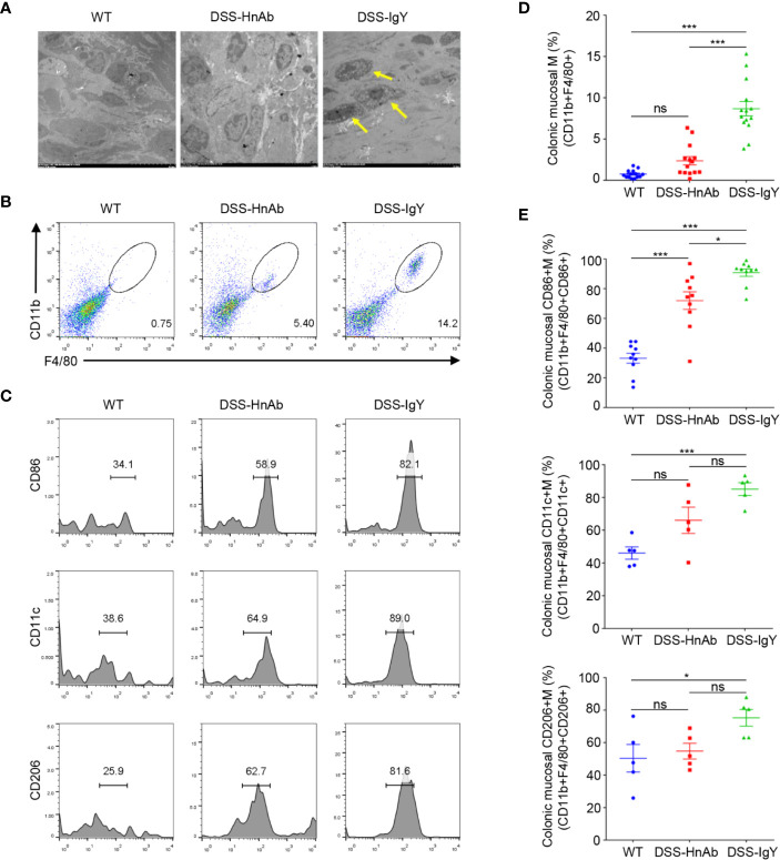
Figure 4.
Macrophages and CD86+ macrophages are decreased in DSS/HnAb-treated colon. (A) Macrophages were observed in transmission electron microscopic images. Few macrophages could be found in lamina propria in the control, while an increased number was present in dextran sulfate sodium (DSS)/IgY con. DSS/HnAb treatment reduced the number of lamina propria macrophages. (B) Macrophages were identified by the high expression of F4/80 and CD11b. (C) Activation markers CD86, CD11c, CD206 were used to count macrophages by FACS analysis. (D) Number of macrophages in % of total counted cells was significantly higher in DSS/IgY treated vs. control colon (P<0.001) as well as vs. DSS/HnAb-treated colon (P<0.001), but not in DSS/HbAg-treated vs. control colon. (E) Significant increase of CD86+ macrophages, but not CD11c+ and CD206 macrophages in DSS/HbAg-treated vs. control colon. Data in (A–C) were representative of five independent experiments. Data in (D, E) were presented as mean ± SEM of five independent experiments. *P < 0.05, ***P < 0.001, ns, not significant, by one-way ANOVA with Tukey’s post-test (D, E).

