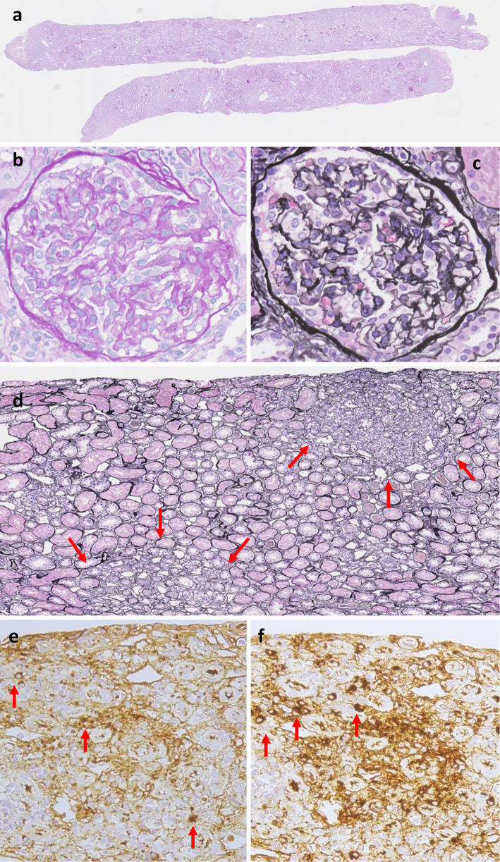
Figure 2.
Histological findings of the kidney. (a) Periodic acid-Schiff (PAS) staining at a low power field (×50). Cortex: medulla ratio was 6: 4. This specimen contained 42 glomeruluses, 11 sclerosed and 2 collapsed. (b) PAS staining of glomerulus at a high power field (×200). There was no endocapillary abnormality. (c) periodic acid-methenamine-silver (PAM) staining of glomerulus at a high power field (×200). There was no subepithelial change. (d) PAM staining of interstitium at low power field (×100). There was segmental lymphoplasmacytic infiltration with fibrosis. (e) IgG staining of interstitium at high power field (×200). (f) IgG4 staining of interstitium (×200) at a high power field.

