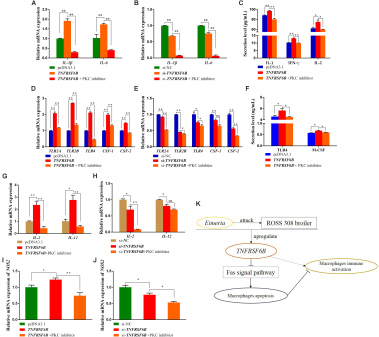
FIGURE 5.
The Apoptosis reversed the TNFRSF6B-mediated strengthening effect on the immune response of macrophages. (A) The mRNA expression of IL-1β and IL-6 after the treatment of TNFRSF6B overexpression and PKC inhibitor. (B) The mRNA expression of IL-1β and IL-6 after the treatment of TNFRSF6B knockdown and PKC inhibitor. (C) The secretion levels of IL-1, IFN-γ, and IL-2 after the treatment of TNFRSF6B overexpression and PKC inhibitor. (D) The mRNA expression of macrophages activation-related genes after the treatment of TNFRSF6B overexpression and PKC inhibitor. (E) The mRNA expression of macrophages activation-related genes after the treatment of TNFRSF6B knockdown and PKC inhibitor. (F) The secretion levels of TLR4 and M-CSF after the treatment of TNFRSF6B overexpression and PKC inhibitor. (G) The mRNA expression of IL-2 and IL-12 after the treatment of TNFRSF6B overexpression and PKC inhibitor. (H) The mRNA expression of IL-2 and IL-12 after the treatment of TNFRSF6B knockdown and PKC inhibitor. (I) The mRNA expression of NOS2 after the treatment of TNFRSF6B overexpression and PKC inhibitor. (J) The mRNA expression of NOS2 after the treatment of TNFRSF6B knockdown and PKC inhibitor. (K) Model of TNFRSF6B-mediated macrophages immune activation. The data was shown as mean ± SEM; *P < 0.05, **P < 0.01, ns, no significant difference.

