Figure 2.
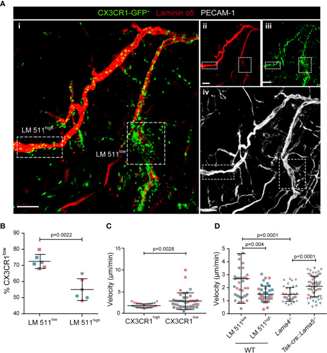
CX3CR1-GFPlow inflammatory monocytes preferentially extravasate at laminin 511low sites. (A) Representative confocal microscopy of whole mount cremaster muscle from a WT host carrying CX3CR1GFP/+ bone marrow stained with anti-laminin α5 and anti-PECAM-1 antibodies (i); single channel images are shown in (ii, iii, iv); scale bar = 100 μm. Experiment was repeated three times with one animal with the same results. (B) To track CX3CR1-GFPlow inflammatory monocytes GFP mean fluorescence intensity (MFI) was measured in situ in areas of high and low laminin 511 expression (see Fig. S2A), revealing a higher proportion of CX3CR1-GFPlow cells, expressed as a percent of total GFP+ cells, at laminin 511low compared to laminin 511high sites; data are means ± SD of three mice from three separate experiments and at least five postcapillary venules in two different areas/mouse; different experiments are marked by different colors. (C) Quantification of GFP MFI and migration speed of individual CX3CR1high and CX3CR1low cells analyzed in three WT hosts from three separate experiments. (D) Average migration velocities of extravasating GFP+ cells at laminin 511 low and high sites in WT controls, and in Lama4−/− and Tek-cre::Lama5−/− chimeras. Data in (C, D) are means ± SD calculated from 39-64 cells analyzed in three mice per chimera (marked by different colors); movies were captured at 4 h post-administration of CCL2. Significance of data in (B–D) were calculated using Mann-Whitney tests since the data did not pass the normality test by D`Agostino & Pearson; the exact p-values are shown in the graph.

