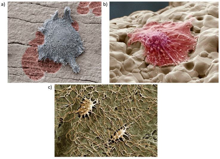
Figure 4.
Coloured scanning electron micrographs of bone cells: (a) Activated osteoclast and resorption pit by kind permission of Timothy Arnett Ref [153]; (b) Osteoblast growing on a bone scaffold made of calcium oxide and silicon dioxide with added strontium and zinc by kind permission of Guocheng Wang from [154]; (c) Osteocytes embedded in the bone matrix with long cytoplasmic extensions reaching into the bone tissue, by kind permission of Kevin Mackenzie. Here, the minerals in the bone have been removed by embedding in resin and etching with perchloric acid. This reveals the spaces in the bone and the shape of the osteocyte cells.

