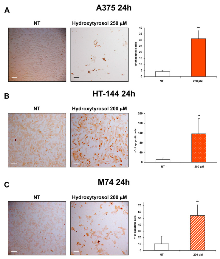
Figure 2.
TUNEL analysis of hydroxytyrosol treated melanoma cells. A375 (A) cells were treated with 250 μM of hydroxytyrosol, HT-144 (B) and M74 (C) cells were treated with 200 μM of hydroxytyrosol for 24 h. Colorimetric TUNEL staining was used to analyse the apoptotic cells of A375 (A), HT-144 (B), and M74 (C) untreated (left panels) and treated (middle panels) cells. Images were acquired with a Zeiss Axioskop 2 Plus microscope using a ×10 objective. Scale bar: 100 μm. The number of stained apoptotic cells were reported in the histograms (right panels). The data are representative of almost three different experiments, the error bars indicate the standard deviation, and statistical significance was analysed by the Student’s t-test: ** p < 0.01 highly significant; *** p < 0.001 very highly significant.

