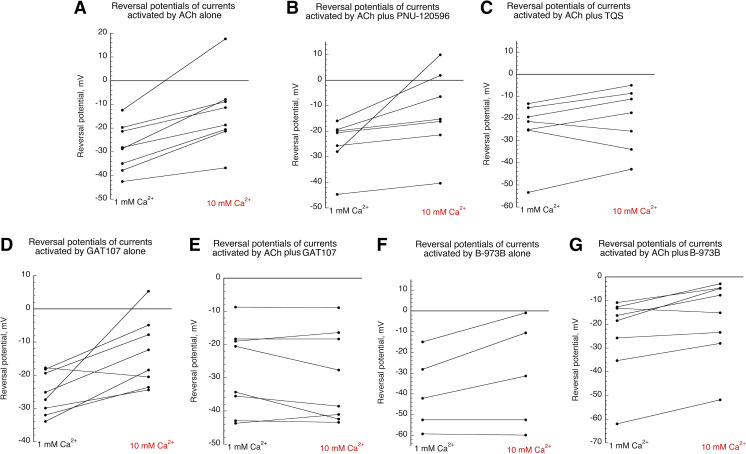
Fig. 10.
Reversal potentials of single cell currents (taken from Fig. 3 and Supplemental Data) plotted for the different conditions. (A) Reversal potentials of currents activated by 30 µM ACh alone in 1 or 10 mM Ca2+ chloride-free Ringer’s solution. (B) Reversal potentials of currents activated by 30 µM ACh coapplied with 10 µM PNU-120596 in 1 or 10 mM Ca2+ chloride-free Ringer’s solution. (C) Reversal potentials of currents activated by 30 µM ACh coapplied with 10 µM TQS in 1 or 10 mM Ca2+ chloride-free Ringer’s solution. (D) Reversal potentials of currents activated by 10 GAT107 applied alone in 1 or 10 mM Ca2+ chloride-free Ringer’s solution. (E) Reversal potentials of currents activated by 30 µM ACh coapplied with 10 µM GAT107 in 1 or 10 mM Ca2+ chloride-free Ringer’s solution. (F) Reversal potentials of currents activated by 10 B-973B applied alone in 1 or 10 mM Ca2+ chloride-free Ringer’s solution. (G) Reversal potentials of currents activated by 30 µM ACh coapplied with 10 µM B-973B in 1 or 10 mM Ca2+ chloride-free Ringer’s solution.

