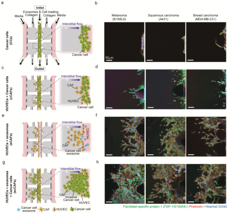
Figure 6.
Three-dimensional microfluidic model for cancer cell invasion. (a,c,e,g) Schematic diagram showing the progression of cancer invasion; (b,d,f,h) confocal images of only cancer cells (CCs) free of human umbilical vein endothelial cells (HUVECs), differentiated cancer-associated fibroblasts (CAFs) induced by cancer cells (cCAFs) or cancer cell-derived exosomes (eCAFs), and cancer invasion created by the exosome-induced CAFs (ecCAFs) from melanoma (B16BL6), squamous carcinoma (A431) and breast carcinoma (MDA-MB-231) cells. This 3D microfluidic model includes an endothelial monolayer composed of HUVECs, CAFs (yellow) induced by cancer cell-derived exosomes (blue) and 3D collagen matrix (gray). Then cancer cells (green) were injected to develop a cancer invasion model. Scale bar: 50 μm.

