Figure 1.
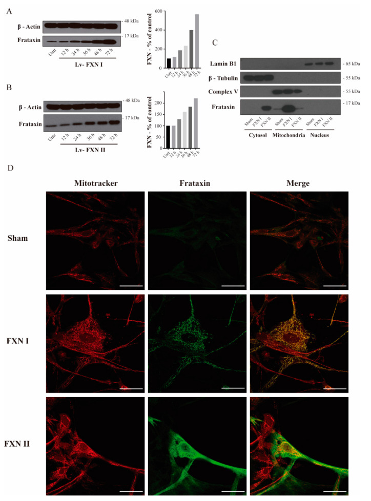
Intracellular localization of frataxin isoform I and II in FRDA-derived OE-MSCs. (A) to (B) OE-MSCs from FRDA patient transduced for 48 h before cell fractionation with either Lv-FXN I (A) or Lv-FXN II (B). (C) Subcellular fractionation of patient-derived OE-MSCs transduced with lentivectors codifying for FXN isoforms. Specific markers were used to identify the appropriate subcellular compartment: Lamin B1 for nucleus, Complex V (ATP-Synthase) for mitochondrion and β-tubulin for cytoplasm. (D) Representative confocal photomicrographs showing FRDA-derived OE-MSCs that were left untreated, or transduced with Lv-FXN I or Lv-FXN II, and stained with Mitotracker (Red) and frataxin (Green). The merged panels show mitochondrial co-localization between frataxin isoform I and Mitotracker, and the cytosolic localization of frataxin isoform II. Calibration bar represents 50 µm.

