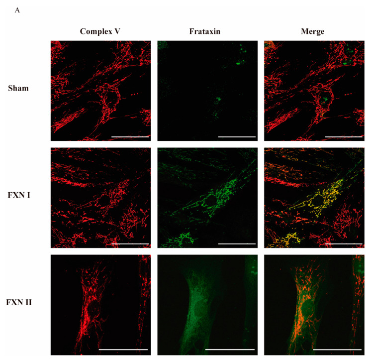
Figure 2.
Intracellular localization of frataxin isoform I and II in fibroblasts from FRDA patients. (A) Representative confocal photomicrographs showing FRDA-derived fibroblasts either untreated, transduced with Lv-FXN I or Lv-FXN II, and stained with antibodies against Complex V (Red) and frataxin (Green). The merged panels show mitochondrial co-localization between frataxin isoform I and Complex V, and the cytosolic localization of frataxin isoform II. Calibration bar represents 50 µm.

