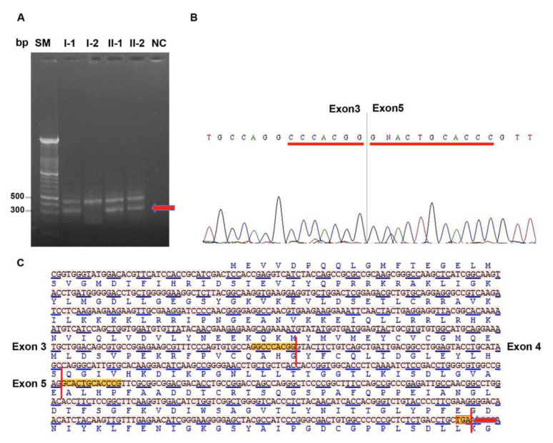
Figure 3.
Identification of the altered splicing isoform. (A) RT-PCR analysis of the cDNA region encompassing exons from 2 to 6. The arrow indicates the abnormal mRNA fragment showing lower molecular weight. (B) Electropherogram showing a STK11 isoform lacking exon 4 and the formation of a new junction between exons 3 and 5. (C) STK11 cDNA sequence in FASTA format showing junctions between exons 3–4 and 4–5; skipping of exon 4 generate a reading frame-shift and formation of a premature stop codon highlighted in the red box and indicated with red arrow. Bp: base pair, SM: size marker, I-1, I-2, II-1 and II-2: subjects of PJS family as reported in pedigree of Figure 2A, NC: RT-PCR negative control without template.

