Figure 5.
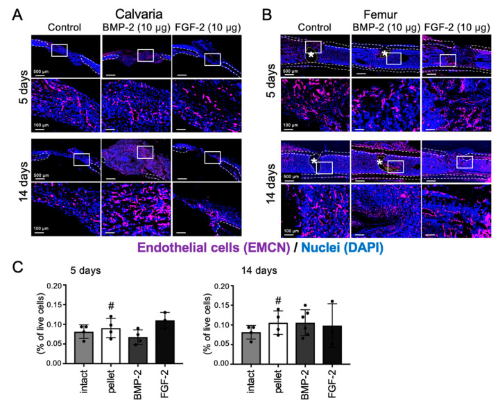
Effects of BMP-2 and FGF-2 on angiogenesis in mouse calvarial and femoral defect. Cross-sectional frozen sections of calvaria (frontal plane, A) and femurs (B) obtained from wild-type mice after 5 and 14 days of collagen pellet implantation. EMCN-positive endothelial cells are shown in purple. Dashed lines indicate the cortical bone (white) and the remained collagen pellet (*, yellow). Nuclei were stained with DAPI (blue). The results are representative of at least three independent experiments. Collagen pellets containing BMP-2 (10 µg) or FGF-2 (10 µg) or DW (control) were transplanted into mouse femur defects and samples were analyzed by FCM (C) at 5 days and 14 days after transplantation. Endothelial cell: CD31⁺CD45⁻. (n = 3–6, One-way ANOVA/Tukey).

