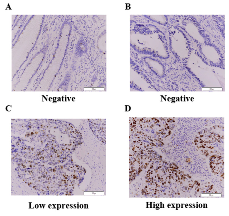
Figure 1.
Immunohistochemistry for HJURP expression in colorectal cancer tissues. (A–D). One hundred and sixty-two CRC samples were stained with HJURP antibody and scored based on both staining intensity and staining frequency. Representative images of tissue slides and immunohistochemistry are shown ((A), Normal negative HJURP expression; (B), Tumor negative; (C), Tumor low expression; (D), Tumor high expression). Original magnification × 200.

