
Figure 7.
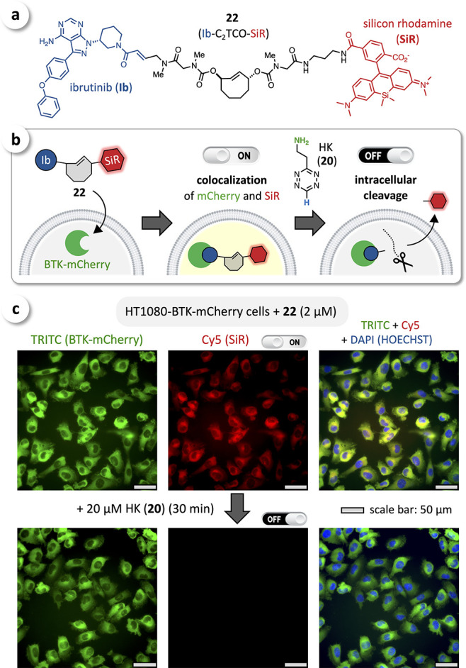
(a) Chemical structure of Ib-C2TCO-SiR (22) as a cleavable fluorescent ibrutinib conjugate. (b) Cellular uptake and binding to BTK of 22 upon treating HT1080-BTK-mCherry cells, and subsequent intracellular cleavage with HK (20). (c) Fluorescence microscopy imaging showed excellent colocalization of mCherry (green) and SiR (red), confirming selective binding of Ib-C2TCO-SiR (22) to BTK (ON-state, top row); efficient intracellular cleavage was observed upon treatment with 20 μM HK (20) for 30 min (OFF-state, bottom row).