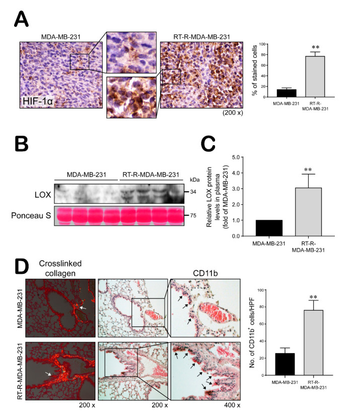
Figure 5.
RT-R-MDA-MB-231 cell-injected mice increased HIF-1α levels and LOX secretion, and premetastatic niche formation. (A) HIF-1α levels detected in tumor section by immunohistochemistry (magnification; ×200 and ×400). Data represent mean values ± SEM (n = 10). ** p < 0.01 compared with the MDA-MB-231-injected mice. (B,C) Secreted LOX levels were detected in the plasma by western blot analysis. Data represent mean values ± SEM (n = 8). ** p < 0.01 compared with the MDA-MB-231-injected mice. (D) Lung tissue sections from MDA-MB-231- and RT-R-MDA-MB-231-injected mice were stained with Picrosirius Red to analyze crosslinked collagen. Recruited CD11b+-immunoreactive bone marrow-derived dendritic cells (BMDCs) near the sites of collagen cross-linkage were stained with anti-CD11b antibody. White arrows indicate representative crosslinked collagen fibers. Black arrows indicate recruited CD11b+ BMDCs (magnification; ×200 and ×400). Data represent mean values ± SEM (n = 10). ** p < 0.01 compared with the MDA-MB-231-injected mice.

