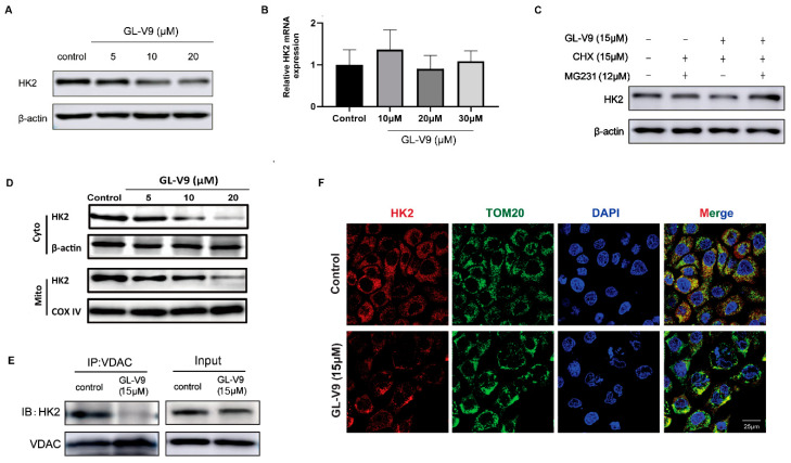
Figure 5.
GL-V9 downregulates protein stability of HK2 and inhibits its mitochondrial location. (A,B) A431 cells were treated with GL-V9 for 36 h. The protein expression (A) and mRNA expression (B) of HK2 were assayed by western blot and RT-PCR, respectively. Bars, SD from three independent experiments. (C) A431 were treated with 15 μM GL-V9 for 24 h, then co-treated with 15μg/mL CHX and 12 μM MG-132 for another 12 h. Then protein level of HK2 was assayed. (D) The expression of HK2 proteins in cytosol and mitochondria was examined by western blot. (E) The binding of HK2 with VDAC was assayed by immunoprecipitation. (F) The location of HK2 in mitochondria was observed by immunofluorescence. TOM20 was a mitochondrial marker. Scale bar = 25 μm.

