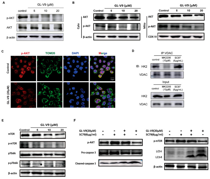
Figure 6.
Inhibition of AKT by GL-V9 is responsibility for the decreasing binding of HK2 in mitochondria and induction of autophagy. (A) A431 cells were treated with GL-V9 for 36 h. The protein expression of AKT and p-AKT were assayed. (B) The expression of AKT and p-AKT proteins in cytosol and mitochondria was examined by western blot. (C) The location of p-AKT in mitochondria was observed by immunofluorescence. Scale bar = 25 μm. (D) A431 cells were treated with 10 μM MK2206 or 6 μg/mL SC97 for 36 h, respectively. The binding of HK2 with VDAC was assayed by immunoprecipitation. (E) Western blot assays were used to examine the protein expression of mTOR, p-mTOR, p70s6k, and p-p70s6k. (F) A431 cells were co-treated with SC79 and GL-V9 for 36 h. The expression of key proteins involved with apoptosis and autophagy were assayed.

