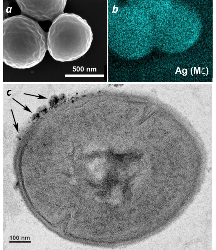
Figure 10.

(a,b) SEM and EDS (Ag Mζ) images of S. aureus after a 30-min exposure to 10-nm GaHb–AgNPs. (c) TEM image of GaHb–AgNPs (indicated with arrows) on the outer bacterial wall (additional images in ESI). TEM sample was prepared using S. aureus exposed to GaHb–AgNPs for 1 hour, then sectioned by microtomy.
