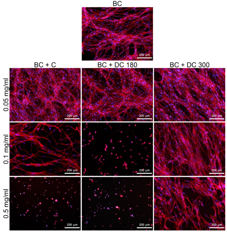
Figure 8.
Morphology of human dermal fibroblasts on pristine bacterial nanocellulose (BC) and on nanocellulose loaded with pure curcumin (BC + C), or with curcumin degraded at 180 °C (BC + DC 180) or at 300 °C (BC + DC 300) at various concentrations (0.05, 0.1, and 0.5 mg/mL) on day 7 after cell seeding. The cells were stained with phalloidin-TRITC (red; F-actin cytoskeleton) and with DAPI (blue; cell nuclei). Olympus IX 51 microscope, obj. 10×, DP 70 digital camera.

