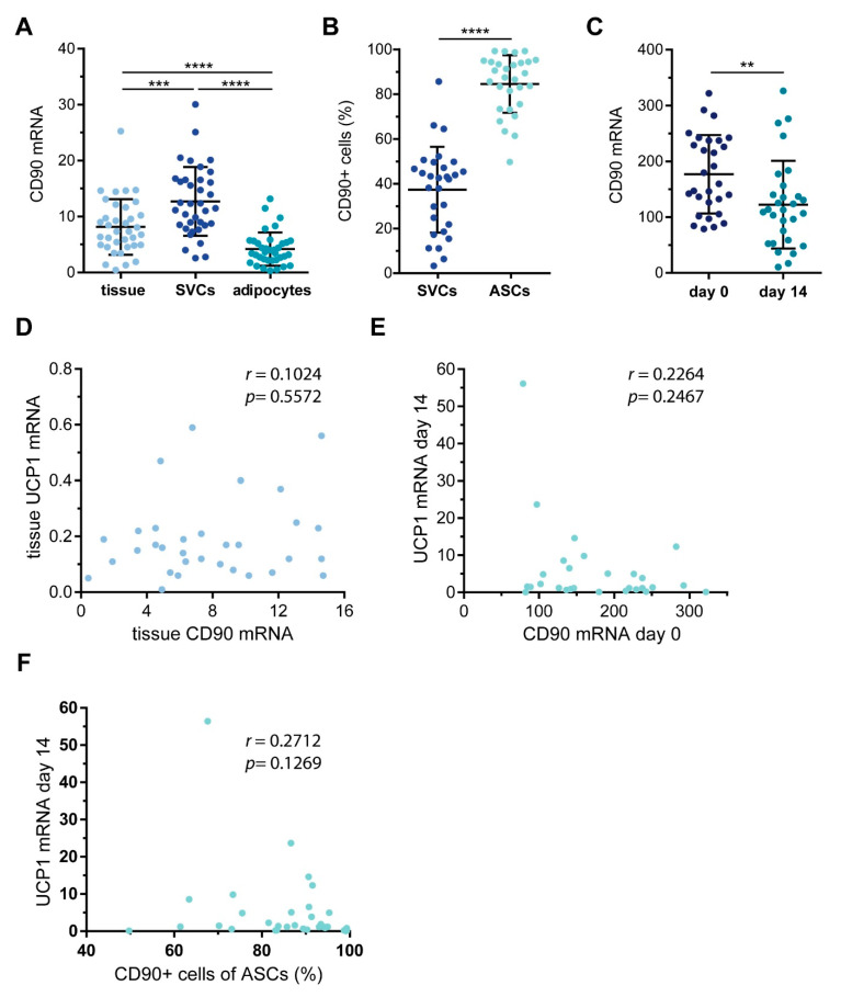
Figure 2.
CD90 expression in white adipose tissue (WAT)-derived SVCs and ASCs. WAT samples were obtained from 29 patients undergoing plastic surgery. (A) The mRNA expression of CD90 was determined by RT-qPCR in whole tissue as well as isolated SVCs and adipocytes. TF2B expression was used to normalize the data. Data are displayed as mean ± SD. *** p < 0.001, **** p < 0.0001, one-way ANOVA with Tukey correction. (B) SVCs and ASCs were analyzed by flow cytometry to quantify the percentage of CD90+ cells. Data are displayed as mean ± SD. **** p < 0.0001, Student’s paired t-test. (C) ASCs taken into culture and subjected to adipogenic differentiation. RNA was isolated before adipogenic induction (day 0) and 14 days after (day 14). The mRNA expression of CD90 was determined by RT-qPCR. TF2B expression was used to normalize the data. Data are displayed as mean ± SD. ** p < 0.01, Student’s paired t-test. (D) Correlation of WAT mRNA expression of UCP1 and CD90. (E) Correlation of ASC mRNA expression of CD90 and corresponding adipocyte mRNA expression of UCP1. (F) Correlation of ASCs percentage of CD90+ cells and corresponding adipocyte mRNA expression of UCP1. Spearman correlation coefficient r and p value are given in each scatter plot.

