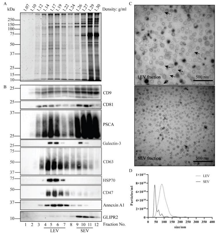
Figure 1.
Isolation and characterization of LEV and SEV. Total EV in seminal plasma of vasectomized men were collected by UC at the interface of a sucrose block gradient. LEV and SEV were separated by their distinct velocities during upward displacement into a continuous sucrose density gradient. (A) Gradient fractions were analyzed by SDS-PAGE followed by Sypro ruby staining for total protein. (B) Gradient fractions were analyzed by immunoblotting for the presence of EV associated proteins, including CD9, CD81, PSCA, Galectin-3, CD63, HSP70, Annexin A1, and GLIPR2. Molecular weight markers are indicated on the left in kDa. (C) Particles in the LEV and SEV containing fractions were analyzed by TEM. Scale bar, 500 nm. Arrows exemplify incidental SEV in the LEV isolate. (D) Size distribution of the particles in LEV and SEV isolates as determined by NTA.

