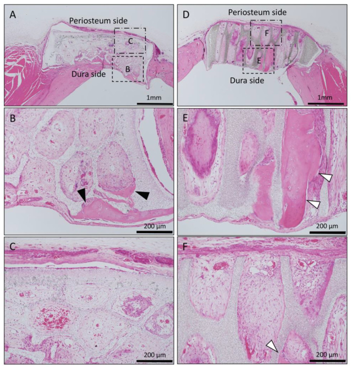
Figure 5.
Histological images of β-TCP with Collagen gel alone at 4 weeks after implantation. (A) Low-magnification image of horizontal β-TCP. No inflammatory granulation tissue was observed around β-TCP. (B,C) Higher-magnification image of corresponding outlined area in (A). Inflammatory cell infiltration was poor in all of the TCP holes, and vascular invasion was observed. Only a small amount of bone tissue was found near the dura (black arrowheads), and the formation of bone tissue was not found in the holes near the periosteal side. (D) Low-magnification image of vertical β-TCP. No inflammatory granulation tissue was observed around β-TCP. (E,F) Higher-magnification image of corresponding outlined area in (D). The formation of bone tissue was observed in some through-holes of the dura side (white arrowheads); however, no bone tissue formation was observed in the periosteum side.

