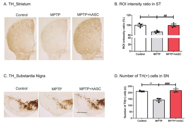
Figure 2.
Effects of hASC transplantation on the loss of tyrosine hydroxylase (TH)-positive dopaminergic neurons in the striatum and substantia nigra of MPTP-induced Parkinson’s disease mice. The brain slices were immunostained using an anti-TH antibody in the striatum and substantia nigra regions. TH-positive dopaminergic neurons in the striatum and substantia nigra were microscopically visualized using diaminobenzidine (DAB) stain. (A) Representative images of TH immunoreactivity in the striatum from mice. Scale bars, 200 μm. (B) The average intensity of TH-positive cells in the striatum was illustrated using a graph. (C) Representative images of TH immunoreactivity in the substantia nigra from mice. Scale bars, 200 μm. (D) The average number of TH-positive cells in the substantia nigra were counted and illustrated using a graph. Dopaminergic neuronal cell death in the striatum and substantia nigra regions sharply increased following the injection of MPTP, and dopaminergic neurons recovered with hASC transplantation. All data are indicated as mean ± standard error (n = 3 per group). * p < 0.05 and ** p < 0.01 vs. Control, and ## p < 0.01, ### p < 0.001 vs. MPTP using one-way ANOVA and Kruskal–Wallis multiple comparisons test.

