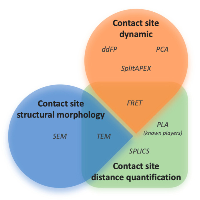
Figure 3.
Three main sub-groups of useful methods to characterize ER–mitochondria contact sites have been identified, which provide information on contact site dynamics, contact site distance, and contact site structural morphology. Methods are clustered on the basis of their utility to output a specific information; at intersections, those amenable approaches able to provide more than one information on contact sites have been reported. Abbreviations: ddFP (dimerization-dependent fluorescent protein) [79], PCA (protein-fragment complementation assay) [81], SplitAPEX (split-ascorbate peroxidase) [104], PLA (proximity ligation assay) [100], SPLICS (split-GFP) [89], TEM (transmission electron microscopy) [105], and SEM (scanning electron microscopy) [111].

