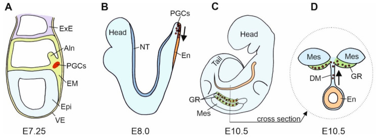
Figure 2.
Scheme of the mouse primordial germ cells (PGCs) fate. (A). In the mouse gastrula at E7.25, E-cadherin positive (+) PGCs are present at the site of their origin, the E-cadherin positive embryonic mesoderm (EM). At this stage, mesodermal cells segregate into a group of E-cadherin positive somatic cells, E-cadherin positive PGCs, and E-cadherin negative (−) cells that form the allantois (Aln). (B). In the mouse neurula at E8.0, PGCs translocate from the mesoderm to the E-cadherin positive endoderm, En (a region of hindgut formation). At this stage, the expression of E-cadherin in PGCs decreases. Subsequently, PGCs migrate individually in the anterior direction. (C) (lateral view) and (D) (cross section). In the mouse embryo at E10.5, PGCs leave the hindgut, increase E-cadherin expression and interconnect by filopodia. PGCs migrate dorsally through dorsal mesentery (DM) to the genital ridges (GR). Epi—epiblast; ExE—extraembryonic ectoderm; Mes—mesonephros; NT—neural tube; VE—visceral endoderm.

