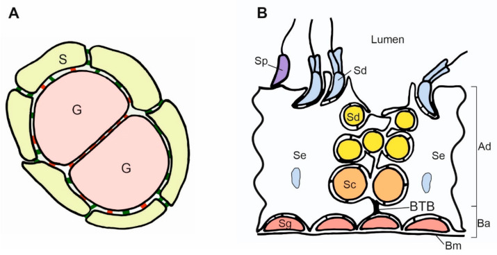
Figure 3.
Scheme of contacts between germ and somatic cells in the gonads. (A). Adjacent germ cells (G) in the nest are connected by E-cadherin (red) and enclosed by the somatic cells (S). The N-cadherin (green) is present between somatic cells, and both E- and N-cadherin are present between somatic and germ cells. (B). Scheme of the mouse seminiferous epithelium. The spermatogonia (Sg) are located in the basal region of the seminiferous epithelium and adhere to the basement membrane (Bm) and Sertoli cells (Se). The accumulation of the adherence junctions forms the blood–testis barrier (BTB) above the spermatogonia. BTB divides the epithelium into two compartments: basal (Ba) and adluminal (Ad). The transitional disassembly of BTB allows pachytene spermatocyte (Sc) passage from the basal to the adluminal compartment. While spermatogenesis progresses, the round and elongated spermatids (Sd) become oriented toward the lumen of the seminiferous tubules. All the germ cells form contacts with Sertoli cells. Spermatids develop flagellum while still attached to the Sertoli cells. In the process of spermiation, the germ cells become released (detached) from the Sertoli cells, and from that moment, they are called the spermatozoa (Sp). The spermatogenesis, including BTB assembly and disassembly, the passage of germ cells toward the lumen, and spermatozoa release, are regulated by changes in cell adhesion.

