Figure 6.
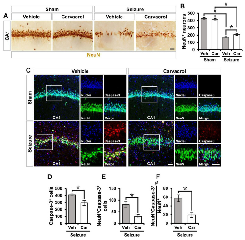
Carvacrol treatment reduces apoptotic neuronal death following pilocarpine-induced SE. (A) Representative images showing the expression of NeuN+ live neurons in the hippocampal CA1 from the vehicle- and carvacrol-treated groups 1 week after sham or SE. Scale bar, 100 µm. (B) Quantification showing the number of NeuN+ neurons as determined in the same hippocampal region (mean ± SEM; n = 6 from each sham group, n = 10 from each seizure group). * p < 0.05 vs. the vehicle-treated group; # p < 0.05 vs. the sham-operated group (Kruskal–Wallis test followed by a Bonferroni post-hoc test; chi square = 23.958, df = 3, p < 0.001). (C) Double immunofluorescent images representing the neuronal marker NeuN+ cells (green) co-labeled with the cleaved caspase-3 (red) in the hippocampal CA1 from the vehicle- and carvacrol-treated groups after SE. The nuclei are counterstained with DAPI (blue). Scale bar, 50 µm. (D–F) Quantification of the number of caspase-3+ (D) and NeuN+Caspase-3+ cells (E) and the percent of NeuN+Capase-3+ cells over total NeuN+ cells (F) as determined in the same hippocampal region (mean ± SEM; n = 4–6 per group). * p < 0.05 vs. the vehicle-treated group (Mann–Whitney U test: Figure 6D: z = 2.082, p = 0.041; Figure 6E: z = 2.242, p = 0.026; Figure 6F: z = 2.882, p = 0.002).

