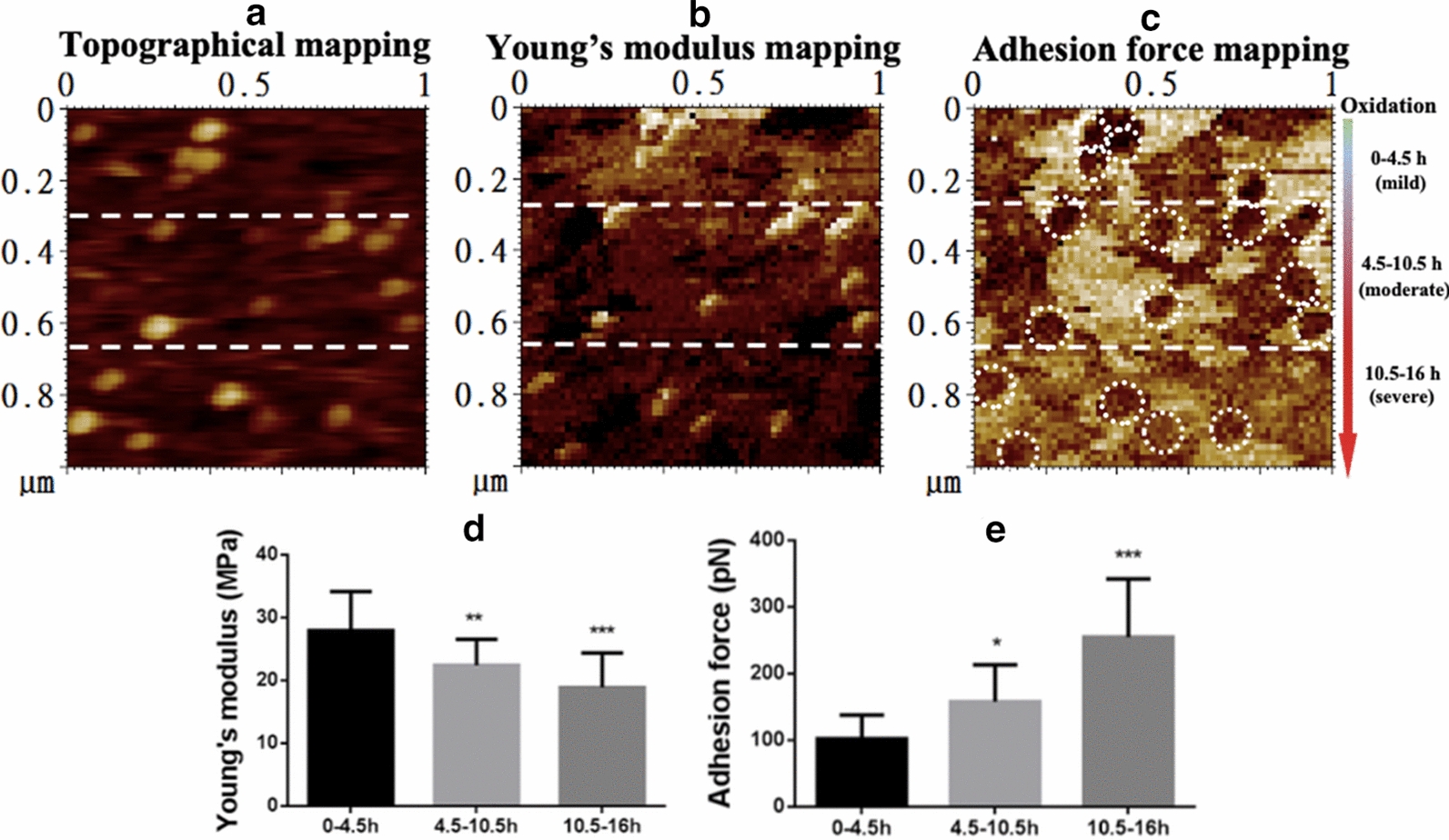
Fig. 4.

Dynamic observation of the effects of oxidation on the biomechanical properties of LDL particles. Native LDL particles pre-immobilized on functionalized mica were mixed with 5 µM copper sulphate on the imaging stage of atomic force microscope and imaged immediately. The LDL particles were oxidized during imaging which lasted for ~ 16 h. It means that the particles from top to bottom in the same image were oxidized to different extents (from mild to severe). For the convenience of quantification, the image was divided into three sections from top to bottom (as indicated by the dashed lines), representing the LDL particles oxidized for 0–4.5 h (no or mildly oxidized), 4.5–10.5 h (moderately oxidized), and 10.5–16 h (severely oxidized), respectively. a Topographical mapping of the LDL particles in a field of 1 µm × 1 µm. b Young’s modulus mapping of the same particles. c Adhesion force mapping of the same particles. The white dashed circles in the adhesion force images indicate individual particle-like patches corresponding to the oxLDL particles at the same locations in the topographical image. d Quantification of the average Young’s modulus of the particles in each section. e Quantification of the average adhesion force of the particles in each section
