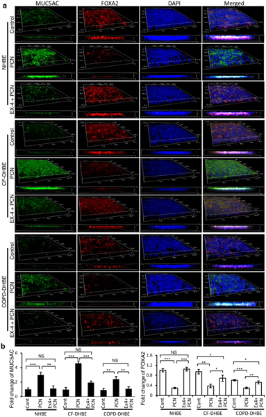
Fig. 4.
Ex-4 restores the expression of FOXA2 to attenuate excessive mucins in pyocyanin-exposed normal (NHBE) and diseased (CF-DHBE and COPD-DHBE) primary airway epithelial cells. Two-week old, partially polarized air-liquid interface (ALI) cultures of airway cells were potentiated to differentiate into mucin secreting goblet cells by exposing to PCN (5 μg/ml) for 24 hours in presence or absence of Ex-4 (1 μg/ml). a MUC5AC and FOXA2 expression were visualized by immunofluorescence labeling using a confocal microscope, and were processed into 3D-models and Z-stack images. Experiments were performed in triplicates independently three times. Representative images from one replicate is shown. b Intensity of MUC5AC and FOXA2 expression was determined using the Z-stack images. Densitometry analyses were normalized against airway cells treated with PBS control (Cont), and mean ± s.e.m from three experiments are shown. MUC5AC and FOXA2 expression were compared by Student’s t-test. *p < 0.05, **p < 0.01, ***p < 0.001. NS: Not significant. MUC5B results are presented in the Fig. S2.

