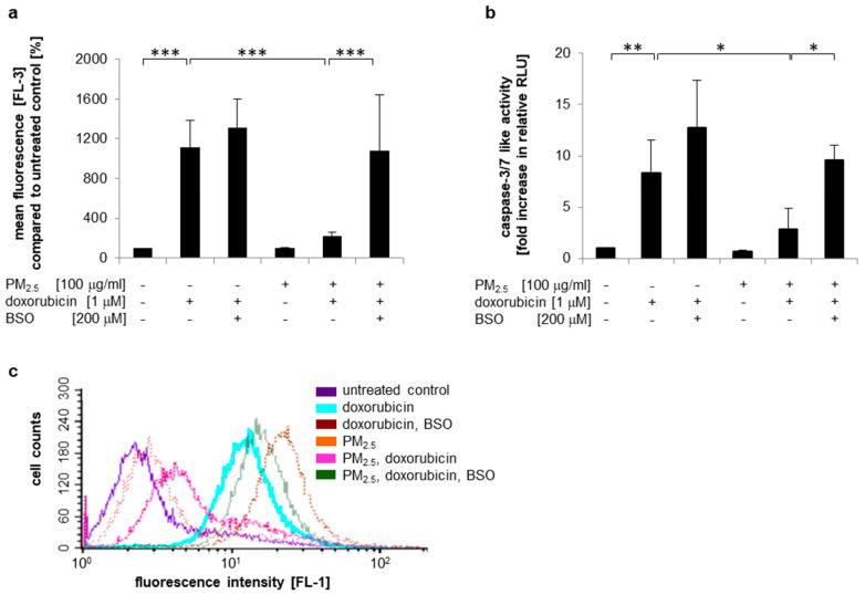
Figure 7.
PM2.5 reduces doxorubicin resistance by a GSH-dependent mechanism. BEAS-2B cells were cultured in the presence of 100 µg/mL PM2.5 for 3 to 5 weeks, before final passage and re-exposure to 100 µg/mL PM2.5 for 48 h. BSO (200 µM) was added 1 h prior to the last PM2.5 exposure, doxorubicin (1 µM) was added 24 h after the last addition of PM2.5. Cells that were never exposed to PM2.5 served as controls. (a) Quantification of the intracellular doxorubicin content by flow cytometry. Mean fluorescence values (FL3) were compared to untreated cells (n = 3). (b) Detection of apoptosis by a caspase-3/7 activity assay (n = 3). (c) Detection of apoptosis by Annexin V staining and flow cytometry. A representative histogram is shown (n = 3). RLU: relative light units. Values are depicted as means and +standard deviations; * p < 0.05; ** p < 0.01; *** p < 0.001.

