
Figure 2.
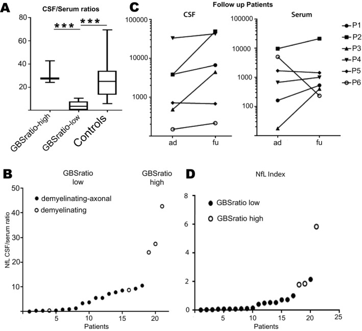
(A) CSF/serum NfL ratio are separated in groups with high and low ratio. High NfL ratios can be found in patients with predominant CNS affection and in control. (B) NfL CSF/serum ratios in all GBS patients, full circle = mixed demyelinating axonal affection in nerve conduction velocity, hollow circle = demyelinating lesions only in nerve conduction velocity. (C) NfL indices of all GBS patients. Full circle meaning NfL‐ratiolow and hollow circle representing NfL‐ratiohigh. (D) CSF‐NfL (left) and Serum NfL (right) levels in follow‐up GBS patients (ad = at diagnose, fu = follow up) Significant difference is marked with *.